Knee Stability
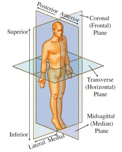
Human Planes [1]
Human Body
The human body is divided up into 3 planes to describe the positions of a body.
Anterior - Posterior
Is defined by the coronal plane as shown. Posterior areas on the body are behind this plane and anterior areas to the front of this plane.
Lateral - Medial
Defined by the Median Plane. Medial describes areas closer to the central plane and lateral away from the body and centre of the plane.
Superior - Inferior
This plane is parallel to the ground and defines the vertical position on the body.
Bone Anatomy
There are 3 main bones that are involved in the knee joint. Starting from the anterior side:
Femur
This is the largest and one of the strongest bones in the body. It translates the forces from the hip joint through to the knee joint. It also acts as a rigid body for muscles that move the knee and the hip joint. The end of the femur has two large condyles that are the articular parts of the femur.
Patella
Links the quadricep muscles over the knee to the tibia. It protects the knee ligaments while the inside is lined with cartilage to reduce friction in the knee. It also increases the pully length of the muscles, increasing the force they can exert in extension.
Tibia
This is the main bone of the calf. The pateller ligament is attached to the front of the tibia along with the other muscles that pass over the bone. It widens at the anterior side on which cartilage sits to cushion the femur.
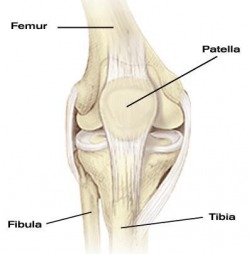
Bones around the knee [2]
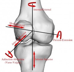
Knee Movements
The knee has 6 degrees of freedom as shown on the right. The main movements of the knee are flexion and extension. The knee also rotates as it locks out at full extension. As the femur reaches the final 30 degrees of extension the femur rotates medially into a locked out position and at this point it is in its most stable position.
Knee flexion is when the angle between the femur and tibia decreases. Knee extension is when the angle increases.
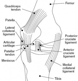
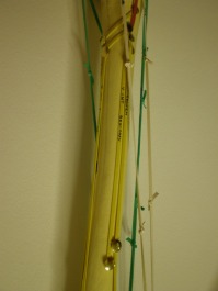
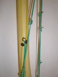
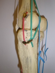
Ligaments (Red Bands)
Ligament are fibrous tissues that connect bones to bones. They are very stiff with very little stretch when put under tension. There are 4 main ligaments around the knee that provide it with stabiltiy.
Cruciate Ligaments
These ligaments connect from the tibial plate to each femur condyle. The cruciate help prevent the tibia moving from the femur in in the anterior-posterior direction. The anterior cruciate ligaments prevents the tibia sliding posteriorly and the posterior cruciate prevents the tibia sliding anteriorly.
Collateral Ligaments
These ligaments go from the medial and lateral side of the femur to the tibia and fibula respectively. These provide strength if the knee is being deflected in the medial-lateral directions.
Muscles Around the Knee
Knee Extension
The main muscle groups that produce knee extension are the quadriceps (yellow bands). As the name suggests there are four muscles in this group.
Rectus Femoris
This muslce sits at sits above the other 3 and originates from the front of the pelvis. It then attaches to the quadricep tendon.
Vastus Medialis
One of three muscles that attach to the femur. This muscle arises from the rear of the femur on the medial side and then attaches to the quardrilateral tendon.
Vastus Lateralis
This muscle also is attached to the posterior side of the femur and wraps round to the quadrilateral tendon.
Vastus Intermedius
This muscle attaches on to anterior and lateral surface of the femur and runs underneath the Rectus Femoris to join the Quadriceps Tendon.
For a complete list of muscle with anatomical origins and insertions a spreadsheet can be downloaded below.
| muscle_insertion_points.pdf |

Knee Flexion
The hamstrings are the main group of muscles that make the knee flex. There are also other muscles that aid flexion which also rotate the knee in the medial lateral directions.
Hamstrings (Green Bands)
Bicep Femoris (Above left)
There are two parts to this muscle, the long head and short head. The long head originates from the rear of the pelvis and the short head from the rear of the femur. They both then come together and attach to the lateral side of leg to the head of the tibia.
Semitendinosus and Semimembranosus (Below Left)
Both attach to the same point as the Bicep Femoris. The Semitendinosus is made of largely of a tendon that then runs to the medial tibial surface. The semimembranosus attaches to the tibial medial condyle.
Gracilis (White)
Attaches to the rear of the pelvis and runs posteriorly and attaches to the medial surface of the tibia.
Sartorius (White)
Attaches to the anterior side of the pevis then passes round the medial side of the femur and attaches to to the same area as the Gracilis and Semitendinosus.
Gastrocnemius (Blue)
Attaches to the ankle then passes up the posterior of the tibia and splits to attach to the medial and lateral side of the femur.
top of page
