Knee Stability
Thesis Work Completed.
Anatomy Laboratory
An anatomy lesson at the Edinburgh University Medical School gave us the chance to observe real bones and knee joints. The geometry of the bones plays a great part in the stability of the knee joint. Things that were hard to imagine from reading were easily shown using the real bones. The medial condyle, which is at the bottom of the femur, is curved as is the tibial plateau which its sits on. This prevents the femur bone from moving medially. The rotation of the knee joint when it “locks” into its fully extended position was seen. This is not a movement that is easily noticed but plays a significant part in the stability of the joint.
The anatomist demonstrated how this along with the collateral ligaments stopped the knee from moving in directions that would damage the knee joint. Using real knee joints the forms that the ligaments take were easy to distinguish, far more than form the literature that had been read. The lateral collateral ligament is a cord like ligament compared to the medial collateral ligament that has a large surface area. The purposes of the cruciate ligaments were easily demonstrated, on the knee joint, in preventing the femur moving forward and backwards on the tibia. A whole leg was used to show us the muscles that are involved in the movement of the knee joint. We were shown in great detail the attachment points of the muscles and shown what part all the muscles play in the flexion, extension and rotation of the knee joint.
3D Model
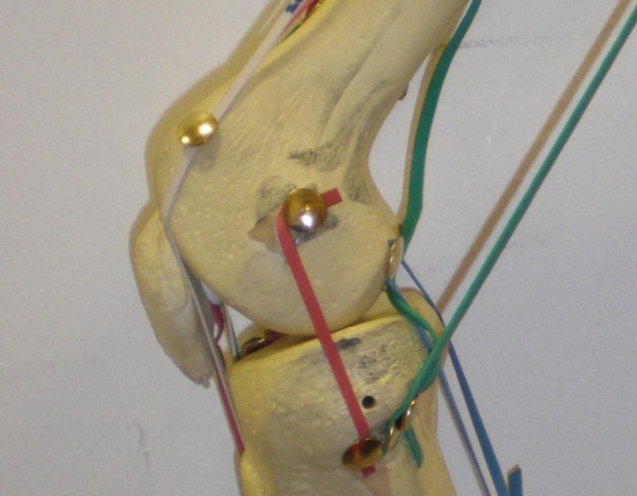
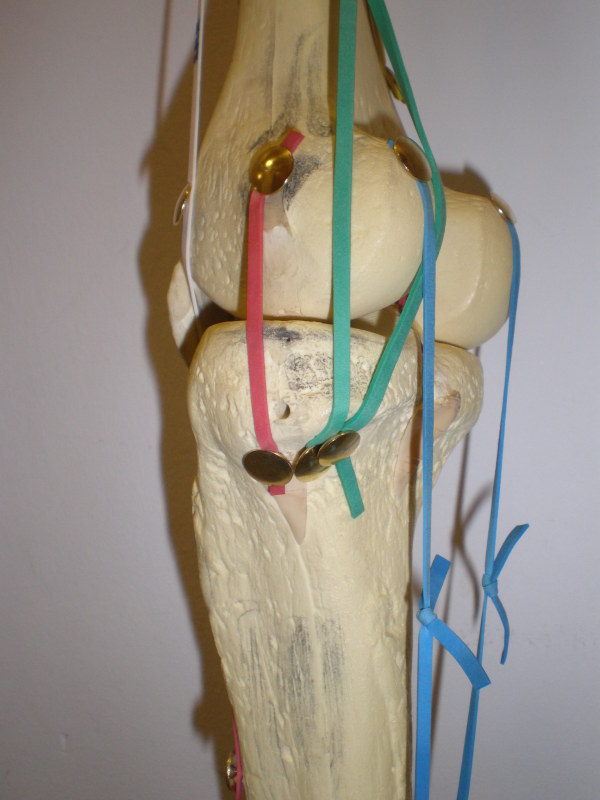
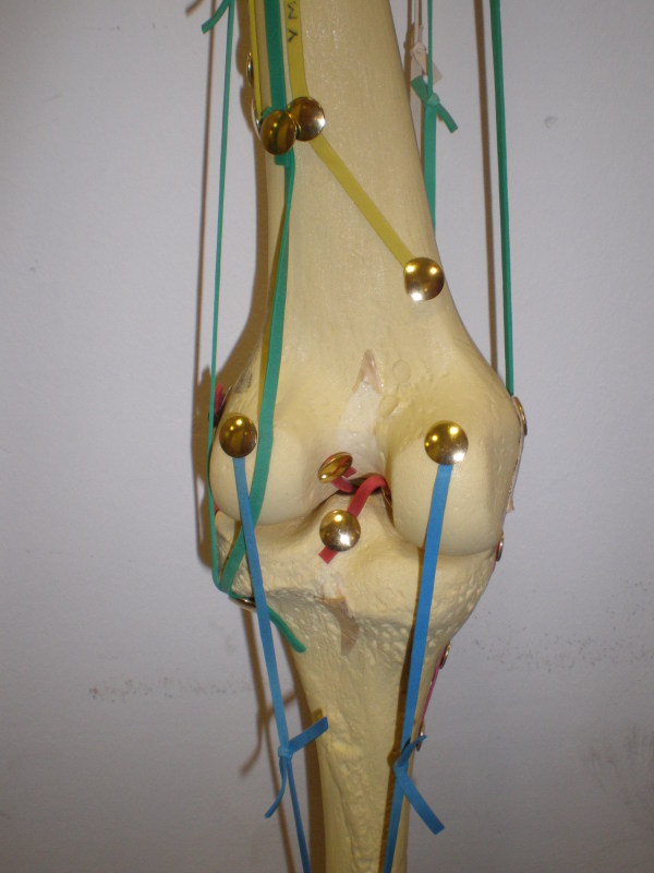
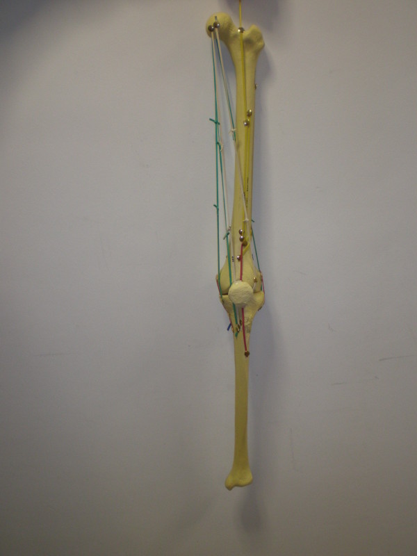
Using the knowledge gained from the anatomy lab, we created a 3D model of the femur and tibia using plastic bones and elastic bands. Muscles and ligaments are represented by elastic bands and are grouped into colours and the key for this can be seen below the images.
Due to the lack of pelvis and fibula in the model, muscles which have connection points on these bones, are represented by being attached to very head of the femur in the case of the pelvis and on the surface where the tibia meets the fibula.
The superficial muscles such as the vastus lateralis were represents by numerous elastic bands, marking out the boundary lines to show the huge surface area that they have. We did not want to mis-represent this muscle especially, as a single line.


Muscle Colour Key
Red – Ligaments Green – Hamstrings Blue – Gastrocnemius White – Sartorius and Gracilis Yellow – Quadriceps
Red – Ligaments Green – Hamstrings Blue – Gastrocnemius White – Sartorius and Gracilis Yellow – Quadriceps
Open Sim Software
Opensim is an open source computer programme that allows the simulation of the musculo skeletal system. We have used this programme to complete tutorials that allowed us to simulate a walking motion with both legs. The model has all the muscles attached and programme allows the user to plot values such as moment arm, muscle length and muscle force against the flexion angle of the knee. The video below shows one of the avaliable models, which we have applied a walking motion to. The Opensim program can be found at http:\simtk.org. The graph shows the forces in the knee flexion muscles during the walking motion.

Graph produced of muscle forces for knee extensor muscles
